Jagannath Gangareddy, Pratyasha Rudra, Manohar Chirumamilla, Sudheer Ganisetti,Subramanian Kasimuthumaniyan, Sourav Sahoo, K. Jayanthi, Jagannath Rathod,Venugopal Rao Soma, Subrata Das, Nitya Nand Gosvami, N. M. Anoop Krishnan,Kjeld Pedersen, Swastik Mondal,* Srabanti Ghosh,* and Amarnath R. Allu
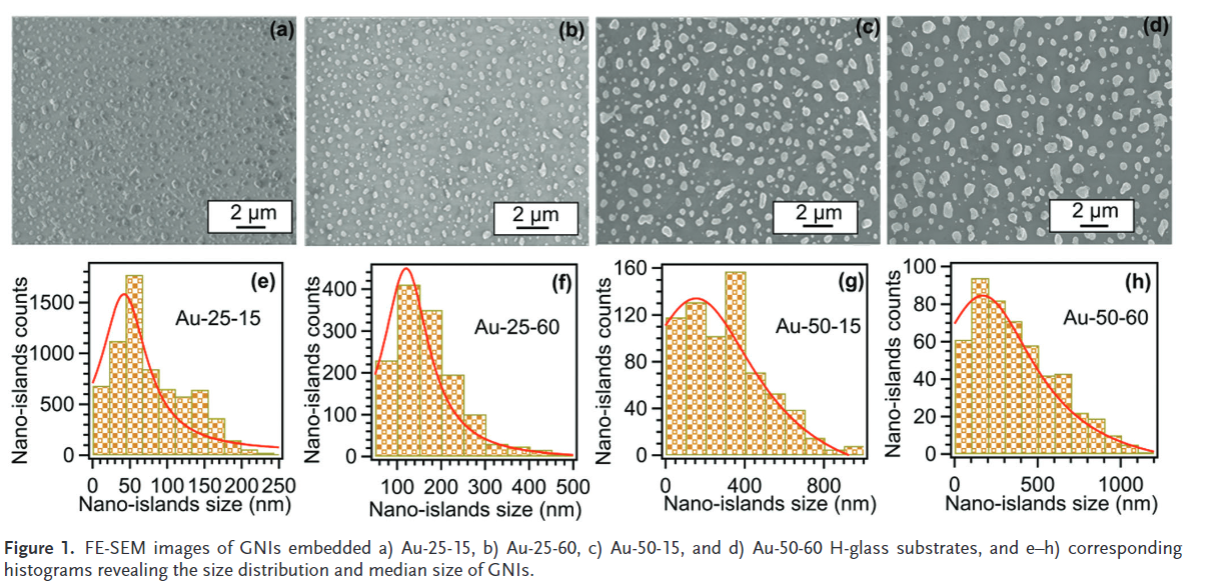
Metal nanoparticles (MNPs) are synthesized using various techniques ondiverse substrates that significantly impact their properties. However, amongthe substrate materials investigated, the major challenge is the stability ofMNPs due to their poor adhesion to the substrate. Herein, it is demonstratedhow a newly developed H-glass can concurrently stabilize plasmonic goldnanoislands (GNIs) and offer multifunctional applications. The GNIs on theH-glass are synthesized using a simple yet, robust thermal dewetting process.The H-glass embedded with GNIs demonstrates versatility in its applications,such as i) acting as a room temperature chemiresistive gas sensor (70%response for NO 2 gas); ii) serving as substrates for surface-enhanced Ramanspectroscopy for the identifications of Nile blue (dye) and picric acid(explosive) analytes down to nanomolar concentrations with enhancementfactors of 4.8 × 10 6 and 6.1 × 10 5 , respectively; and iii) functioning as anonlinear optical saturable absorber with a saturation intensity of 18.36 ×10 15 W m−2 at 600 nm, and the performance characteristics are on par withthose of materials reported in the existing literature. This work establishes afacile strategy to develop advanced materials by depositing metal nanoislandson glass for various functional applications.
Keywords: glasses, gold nanoislands, NO 2 gas sensors, saturable absorber, surface-enhanced Raman spectroscopy substrate, stability of gold nano-islands