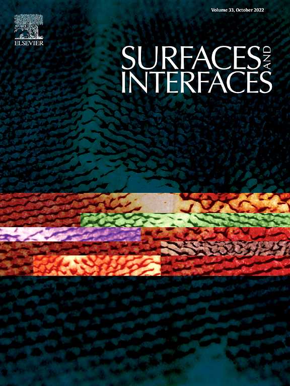
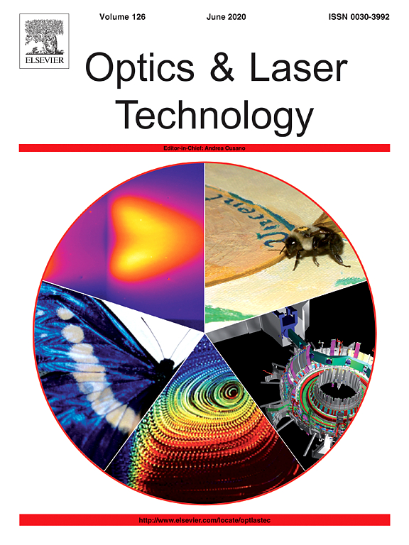
SERSitive substrates in publications

Mocci F, Olla C, Cappai A, Corpino R, Ricci PC, Chiriu D, Salis M, Carbonaro CM.
The molecular model is one of the most appealing to explain the peculiar optical properties of Carbon nanodots (CNDs) and was proven to be successful for the bottom up synthesis, where a few molecules were recognized. Among the others, citrazinic acid is relevant for the synthesis of citric acid-based CNDs. Here we report a combined experimental and computational approach to discuss the formation of different protonated and deprotonated species of citrazinic acid and their contribution to vibrational and magnetic spectra. By computing the free energy formation in water solution, we selected the most favoured species and we retrieved their presence in the experimental surface enhanced Raman spectra. As well, the chemical shifts are discussed in terms of tautomers and rotamers of most favoured species. The expected formation of protonated and de-protonated citrazinic acid ions under extreme pH conditions was proven by evaluating specific interactions with H2SO4 and NaOH molecules. The reported results confirm that the presence of citrazinic acid and its ionic forms should be considered in the interpretation of the spectroscopic features of CNDs.
Keywords: SERS, citrazinic acid; Raman spectroscopy; NMR spectroscopy; carbon nanodots; DFT calculations
Antonio Cappai, Claudio Melis, Luigi Stagi, Pier C. Ricci, Francesca Mocci, and Carlo M. Carbonaro
The molecular emission model is the most accredited one to explain the emission properties of carbon dots (CDs) in a low-temperature bottom-up synthesis approach. In the case of citric acid and urea, the formation of a citrazinic acid (CZA) single monomer and oligomers is expected to affect the optical properties of the CDs. It is therefore mandatory to elucidate the possible role of weak bonding interactions in determining the UV absorption spectrum of some molecular aggregates of CZA. Although this carboxylic acid is largely exploited in the synthesis of luminescent CDs, a full understanding of its role in determining the final emission spectra of the produced CDs is still very far to be achieved. To this aim, by relying on purely first-principles density functional theory calculations combined with experimental optical characterization, we built and checked the stability of some molecular aggregates, which could possibly arise from the formation of oligomers of CZA, mainly dimers, trimers, and some selected tetramers. The computed vibrational fingerprint of the formation of aggregates is confirmed by surface-enhanced Raman spectroscopy. The comparison of experimental data with calculated UV absorption spectra showed a clear impact of the final morphology of the aggregates on the position of the main peaks in the UV spectra, with particular regard to the 340 nm peak associated with n-π* transition.
Keywords: SERS, Citrazinic Acid in Aqueous Solution, carbon dots
Malwina Liszewska, Bartosz Bartosewicz, Bogusław Budner, Barbara Nasiłowska, Mateusz Szala, Jan L. Weyher, Igor Dzięcielewski, Zygmunt Mierczyk, Bartłomiej J. Jankiewicz
Raman spectroscopy has become an essential analytical technique for field detection and identification of illicit or dangerous materials such as explosives, but its main drawback is low signal intensity. This problem can be circumvented by using surface enhanced Raman spectroscopy (SERS), in which scattering signals increase significantly for analytes adsorbed onto or near nano structured surfaces of the plasmonic materials. However,despite numerous studies, SERS has still not been widely used in real-world applications. The main goal of the studies describe herein was to investigate the possibility of detection of trace amounts of selected explosive materials on various commercial and non-commercial SERS substrates using portable Raman instruments. Our studies have shown that while portable systems suitable for SERS measurement of trace amounts of explosives are readily available, the problem remains in the selection of reliable and reproducible SERS substrates. Among five investigated SERS substrates only two, Klarite 312 and GaN-pillars allowed for trace analysis of all studied explosive materials. In both cases, detected concentrations of explosives ranged from single to hundreds of μg/cm2 depending on the explosive material and the Raman spectrometer used. Based on our findings, it could be concluded that the best SERS substrates for trace analysis of explosives are substrates with hot spots densely and evenly distributed over a whole active area of SERS substrate
Keywords: Explosives, SERS, SERS substrates, Portable Raman spectroscopy, Trace detection
Byram Chandu, Moram Sree Satya Bharati, Paweł Albrycht, Soma Venugopal Rao
Over the last decade several research groups have accomplished the fabrication of 2D periodic and 3D nanocage like structures on different materials using diverse lithographic approaches. Herein, we present the detailed studies on the fabrication of femtosecond (fs) laser‐induced periodic/ripple‐like surface structures on nickel (Ni) substrate in distilled water whereas 3D-like (nanocages) features on Ni substrates in acetone by tailoring the laser processing parameters (pulse energy). The morphological studies of simultaneously obtained Ni nanoparticles (NPs)/nanostructures (NSs) in distilled water/acetone were meticulously studied using transmission electron microscope (TEM) and field emission scanning electron microscope (FESEM). The fabricated Ni periodic/3D-like structures were gold (Au) plated using thermal evaporation technique and subsequently utilized as surface enhanced Raman scattering (SERS) active sensors for detecting the traces of various analyte molecules such as malachite green (MG) and Nile blue (NB). The grooved Ni-Au substrates allowed us to detect extremely low concentrations of MG (500 pM) and NB (5 nM) and, significantly, utilizing a simple, portable Raman spectrometer. Moreover, the substrates have demonstrated superior reproducibility as well as multi-utility nature with a relative standard deviation (RSD) of <17%. Additionally, Au- coated Ni grooved SERS substrates have demonstrated superior sensitivity and reproducibility in comparison to commercially available Ag-based SERS sensors (SERSitive, Poland). The proposed method of fabricating ripple and nanocages of Ni SERS platforms are highly viable to overcome the cost and one-time usage of substrates for on-site detection of several analyte molecules using a portable/hand-held Raman spectrometer.
Keywords: Laser Induced Periodic Surface Structures (LIPSS), Malachite green, Nanocages, Nickel Nanostructure (NS), Nile blue, SERS, Substrates, Surface scattering, Laser ablation, Raman, Relative standard deviations
Łukasz Richter, Paweł Albrycht, Monika Księżopolska-Gocalska, Ewa Poboży, Robert Bachlińskic, Volodymyr Sashuk, Jan Paczesny, Robert Hołyst
The majority of analytical chemistry methods requires presence of target molecules directly at a sensing surface. Diffusion of analyte from the bulk towards the sensing layer is random and might be extremely lengthy, especially in case of low concentration of molecules to be detected. Thus, even the most sensitive transducer and the most selective sensing layer are limited by the efficiency of deposition of molecules on sensing surfaces. However, rapid development of new sensing technologies is rarely accompanied by new protocols for analyte deposition. To bridge this gap, we propose a method for fast and efficient deposition of variety of molecules (e.g. proteins, dyes, drugs, biomarkers, amino acids) based on application of the alternating electric field. We show the dependence between frequency of the applied electric field, the intensity of the surface enhanced Raman spectroscopy (SERS) signal and the mobility of the studied analyte. Such correlation allows for a priori selection of parameters for any desired compound without additional optimization. Thanks to the application of the electric field, we improve SERS technique by decrease of time of deposition from 20 h to 5 min, and, at the same time, reduction of the required sample volume from 2 ml to 50 μl. Our method might be paired with number of analytical methods, as it allows for deposition of molecules on any conductive surface, or a conductive surface covered with dielectric layer.
Keywords: Deposition, Electric field, Surface-enhanced Raman spectroscopy, SERS, Analyte, Detection
Dávid J. Palásti, Paweł Albrycht, Patrick Janovszky, Karolina Paszkowska, Zsolt Geretovszky and Gábor Galbács
An assessment of the feasibility of using modified surface enhanced Raman scattering substrates (Ag nanoparticles on indium‑tin-oxide glass) for quantitative nanoparticle-enhanced laser induced breakdown spectroscopy (NELIBS) was carried out. Substrates were prepared with different surface coverage from various nanoparticle sizes, and their laser ablation behaviour was tested in detail. It was found that use of those combinations are most beneficial in terms of the signal enhancement factor, which provide the shortest interparticle distances. With the application of 266 nm laser wavelength, long (ms-range) gate width, and optimized laser pulse energy, the best NELIBS signal enhancement was found to be about a factor of three. By using liquid sample deposition by spraying, which was found to provide an even distribution of liquid samples on the substrate surface, successful calibration for Mn, Zn and Cr was performed. The NELIBS signal repeatability from five repeated measurements was found to be comparable to that of LIBS (5–10% RSD). These observations indicate that the NELIBS signal enhancement approach can be used in quantitative analytical applications for liquid samples, if i) the substrate fabrication procedure has good repeatability, ii) surface coverage and nanoparticle size is tightly controlled, iii) a homogenous liquid sample deposition is achieved.
Keywords: Laser induced breakdown spectroscopy (LIBS); Nanoparticle enhanced LIBS (NELIBS); Silver nanoparticles; Indium‑tin-oxide (ITO) glass; SERS
Mss Bharathi, Abdul Kalam, Chandu Byram, Syed Hamad and Venugopal Rao Soma
The development of recyclable surface enhanced Raman scattering (SERS) based sensors has been in huge demand for trace level explosives detection. A simple, hybrid Silicon (Si) nanotextured target-based SERS platform is fabricated through patterning micro square arrays (MSA) on Si using femtosecond (fs) laser ablation technique at different fluences. Using the hybrid target Si MSA substrate loaded/decorated with Ag-Au alloy NPs (obtained using femtosecond ablation in liquids) we demonstrate the trace level detection of organic nitro-explosives [picric acid (PA), 2,4-dinitrotoluene (DNT), and 1, 3, 5-trinitroperhydro-1, 3, 5-triazine (RDX)] and their mixtures. The microstructures/nanostructures of MSA fabricated at an input fluence of 9.55 J/cm2, and decorated with Ag-Au alloy NPs, exhibited exceptional SERS enhancement factors (EFs) up to ∼1010 for MB, ∼106 for PA, and ∼104 for RDX with the detection limits obtained being ∼5 pM, ∼36 nM, and ∼400 nM for MB, PA and RDX respectively. Furthermore, we demonstrate these SERS substrates possess good reproducibility (RSD values < 15%) and a superior performance compared to a commercial Ag substrate (SERSitive, Poland). Three binary mixtures, i.e. MB-PA, MB-DNT, PA-DNT at different concentrations, were also investigated using the same SERS substrate to test the efficacy. Further, the SERS spectra of dyes, explosives, and complex mixtures were utilized for discrimination/classification using principal component analysis.
Keywords: Surface-enhanced Raman scattering, Femtosecond laser ablation, Ag–Au alloy NPs, Methylene blue, Explosives detection
Hrvoje Gebavi, Vlatko Gašparić, Dubravko Risović, Nikola Baran, Paweł Henryk Albrycht and Mile Ivanda
The paper reports on the features and advantages of horizontally oriented flexible silicon nanowires (SiNWs) substrates for surface-enhanced Raman spectroscopy (SERS) applications. The novel SERS substrates are described in detail considering three main aspects. First, the key synthesis parameters for the flexible nanostructure SERS substrates were optimized. It is shown that fabrication temperature and metal-plating duration significantly influence the flexibility of the SiNWs and, consequently, determine the SERS enhancement. Second, it is demonstrated how the immersion in a liquid followed by drying results in the formation of SiNWs bundles influencing the surface morphology. The morphology changes were described by fractal dimension and lacunar analyses and correlated with the duration of Ag plating and SERS measurements. SERS examination showed the optimal intensity values for SiNWs thickness values of 60–100 nm. That is, when the flexibility of the self-assembly SiNWs allowed hot spots occurrence. Finally, the test with 4-mercaptophenylboronic acid showed excellent SERS performance of the flexible, horizontally oriented SiNWs in comparison with several other commercially available substrates.
Keywords: flexible hot spots; horizontal silicon nanowires; 4-mercaptophenylboronic acid; surface-enhanced Raman spectroscopy (SERS); vapour–liquid–solid
Evelin Witkowska, Dorota Korsak, Aneta Kowalska, Monika Księżopolska-Gocalska, Joanna Niedziółka-Jönsson, Ewa Roźniecka, Weronika Michałowicz, Paweł Albrycht, Marta Podrażka, Robert Hołyst, Jacek Waluk & Agnieszka Kamińska
We show that surface-enhanced Raman spectroscopy (SERS) coupled with principal component analysis (PCA) can serve as a fast, reliable, and easy method for detection and identification of food-borne bacteria, namely Salmonella spp., Listeria monocytogenes, and Cronobacter spp., in different types of food matrices (salmon, eggs, powdered infant formula milk, mixed herbs, respectively). The main aim of this work was to introduce the SERS technique into three ISO (6579:2002; 11290–1:1996/A1:2004; 22964:2006) standard procedures required for detection of these bacteria in food. Our study demonstrates that the SERS technique is effective in distinguishing very closely related bacteria within a genus grown on solid and liquid media. The advantages of the proposed ISO-SERS method for bacteria identification include simplicity and reduced time of analysis, from almost 144 h required by standard methods to 48 h for the SERS-based approach. Additionally, PCA allows one to perform statistical classification of studied bacteria and to identify the spectrum of an unknown sample. Calculated first and second principal components (PC-1, PC-2) account for 96, 98, and 90% of total variance in the spectra and enable one to identify the Salmonella spp., L. monocytogenes, and Cronobacter spp., respectively. Moreover, the presented study demonstrates the excellent possibility for simultaneous detection of analyzed food-borne bacteria in one sample test (98% of PC-1 and PC-2) with a goal of splitting the data set into three separated clusters corresponding to the three studied bacteria species. The studies described in this paper suggest that SERS represents an alternative to standard microorganism diagnostic procedures.
Keywords: Salmonella Typhimurium, SERS, ISO methods, Food, Bacteria detection, PCA
Evelin Witkowska, Tomasz Jagielski, Agnieszka Kamińska, Aneta Kowalska, Anita Hryncewicz-Gwóźdź and Jacek Waluk
This paper demonstrates that surface-enhanced Raman spectroscopy (SERS) coupled with principal component analysis (PCA) can serve as a fast and reliable technique for the detection and identification of human fungal pathogens, such as Trichophyton rubrum, Candida krusei, Scopulariopsis brumptii, and Aspergillus flavus. Fungal infections have become one of the leading infectious causes of morbidity and mortality among hospitalized patients and/or immunocompromised hosts. Hence, there is a strong need for the development of new technologies allowing for fast and reliable diagnosis of fungal diseases. Our study shows that the SERS technique effectively distinguishes between selected common fungal pathogens and thus offers taxonomic affiliation of fungi within several minutes. Additionally, the PCA analysis allows performing statistical classification of fungal pathogens studied and identifying the fungal spectrum directly from a clinical sample. Calculated two principal components (PCs) (PC-1, PC-2) are the most diagnostically significant, explain 97% of the variability and enable, with very high probability, discrimination between the four mentioned fungal species. Moreover, the results of this study demonstrate the excellent possibility for the identification of fungi from human skin samples. The research presented in this paper offers an alternative for conventional fungal diagnostics and paves the way for the development of a new, fast, robust, and cost-effective diagnostic test for the detection and identification of fungal pathogens.
Keywords: SERS, Trichophyton rubrum, Candida krusei, Scopulariopsis brumptii, Aspergillus flavus, Bungi detection